What Hoffa's fat pad is, and why the name matters
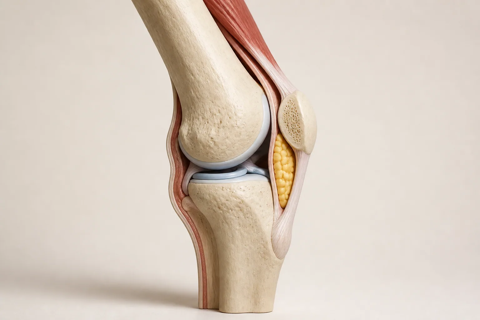
The infrapatellar fat pad, often called Hoffa's fat pad, is a normal soft-tissue structure at the front of the knee. It lies behind the patellar tendon, below the patella, and in front of deeper joint structures. It is inside the joint capsule but outside the synovial cavity. Its shape changes as the knee moves, fitting into the available space around the joint.
Calling it a fat pad can make it sound like spare packing material. It is not. Anatomical research has identified nerve fibers within the pad and adjacent tissue, helping explain why irritation can be painful. Its presence is normal, and the word fat does not imply a weight problem or a need to remove it. Body size cannot establish whether this structure is responsible for knee symptoms.
Impingement describes the possibility of tissue being repeatedly compressed or caught between nearby structures during movement. In practice, clinicians may use terms such as fat pad irritation, Hoffa syndrome, or infrapatellar fat pad impingement with different degrees of certainty. Ask whether the term represents a working clinical explanation, an imaging observation, or a firmly supported diagnosis.
Symptoms may suggest a pattern, but they do not confirm the source
People with suspected fat pad irritation may describe an ache, sensitivity, or pinching sensation near the lower part of the kneecap, sometimes to either side of the patellar tendon. Certain movements or prolonged positions may be more uncomfortable than others. Some notice symptoms as the knee approaches full straightening or rests into extra backward extension.
These descriptions are not unique to Hoffa's fat pad. Nearby tendon, joint, bursal, and other soft-tissue problems can produce overlapping symptoms. Even a very tender spot does not prove which tissue is generating pain. Experimental research shows that stimulation of the fat pad can produce anterior knee pain, but a laboratory pain model is not a clinical test and cannot identify the cause of your symptoms.
The useful information is the pattern: what started it, what changes it, whether the knee swells, and whether movement is genuinely restricted. Record ordinary activities that already reveal the problem, such as standing or walking. There is no need to repeatedly reproduce the pain by pressing into the area or snapping the knee straight.
| Observation | Why it is useful | Why it is not a diagnosis |
|---|---|---|
| Pain near the lower kneecap | Helps locate the symptom for examination | Several structures share this small region |
| Discomfort at the end of straightening | Identifies a movement worth reviewing | More than one condition can limit or hurt during extension |
| Symptoms after a new activity | Suggests a change in exposure or demand | Timing does not establish which tissue is affected |
| Fat pad edema on MRI | May be relevant alongside examination findings | Similar signal changes can occur without clinical impingement |
Why clinicians are cautious about the diagnosis
NHS Tayside's referral guidance explicitly describes fat pad impingement as a controversial diagnosis. It highlights overlap with proximal patellar tendon problems and the possibility of incidental MRI findings. That does not mean your pain is imaginary. It means the available tests do not always identify one clean explanation, so the label should remain open to review.
A reasonable assessment considers the full knee, not just the area mentioned in a report. The clinician may ask about a direct blow, repeated end-range movement, recent surgery, sporting changes, and other symptoms. They may examine movement, swelling, tenderness, and functional tasks as appropriate. The examination should be adapted if the knee is acutely painful or injured.
If the initial explanation remains uncertain, the plan can still be useful. It can identify activities to modify, set an appropriate starting point for rehabilitation, and establish what improvement or deterioration would mean. Diagnostic uncertainty is a reason for careful follow-up, not a reason to cycle through every advertised treatment.
Hoffa fat pad edema is an MRI finding, not a verdict
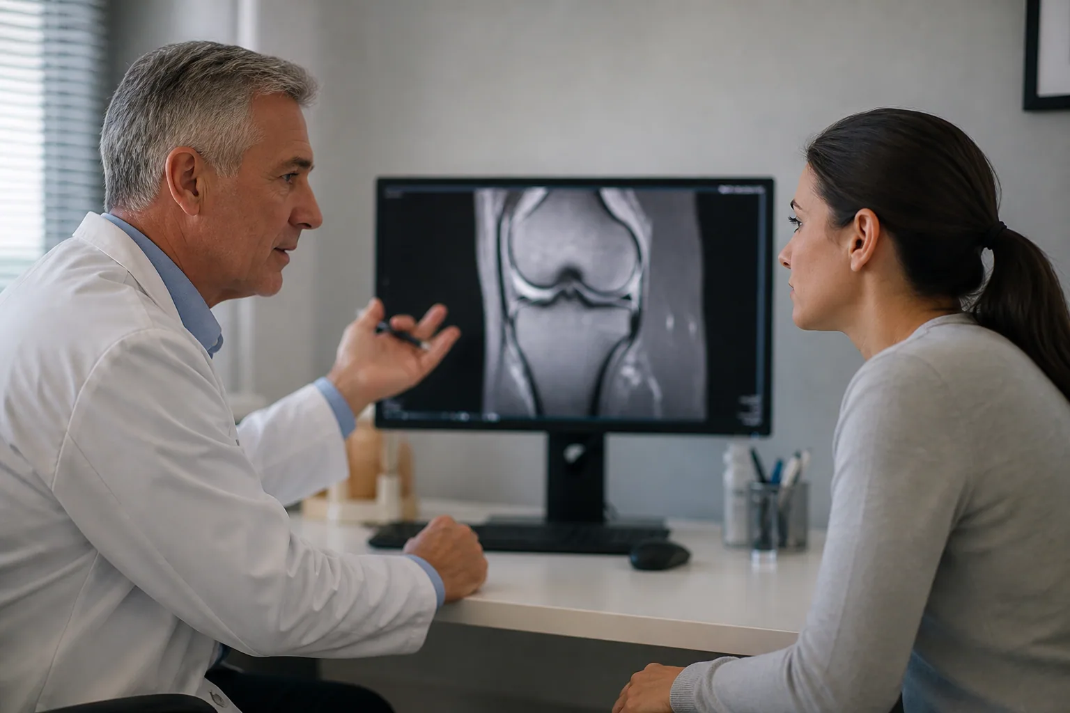
Edema refers to increased fluid-related signal in tissue on suitable MRI sequences. A radiology report may specify the superolateral part of Hoffa's fat pad, meaning the upper outer portion. This precision describes where the signal change appears. It does not establish that the signal is the source of pain, how severe the symptoms should be, or which treatment will work.
In a study comparing MRI appearances with clinical findings, fat pad edema was also present in patients without symptoms of fat pad impingement. The practical lesson is not to ignore the scan. It is to ask whether the scan finding matches the symptom history and examination, and whether other findings better explain the problem. A report should support a clinical decision, not replace one.
Conversely, a person can have genuine pain without a dramatic abnormality on imaging. The intensity of discomfort is not a direct ruler for the size or brightness of an MRI finding. Avoid comparing screenshots online and trying to rank your knee against someone else's. Different scan sequences, positions, and clinical circumstances make those comparisons unreliable.
Patella alta and tracking language need context too
Some reports mention patella alta, a higher-positioned kneecap, or alignment measures related to the patellofemoral joint. Research has found associations between selected extensor-mechanism features and superolateral fat pad edema. These studies examine relationships between measurements. They do not prove that one feature caused an individual's pain or that changing it will necessarily relieve symptoms.
It is easy to move from the phrase maltracking to the belief that the kneecap must be manually put back in the right place. That is an oversimplification. A clinician considers the person's symptoms, function, anatomy, and other findings. Not every anatomical variation needs correction, and a tape pattern or exercise cannot be promised to permanently reposition a kneecap.
If pain is mainly around or behind the kneecap during loaded activities, our pain behind the kneecap guide provides a broader discussion of patellofemoral patterns. The purpose is to understand alternatives and overlap, not to replace one self-diagnosis with another.
Other problems can occupy the same small area
The patellar tendon connects the kneecap to the upper shin. Tendon-related pain may be associated with jumping, running, or other loading, and can overlap the region attributed to the fat pad. Patellofemoral pain can also be felt around the front of the knee. Superficial swelling over or below the kneecap raises a different set of questions from deep discomfort.
A history of a twist, a substantial effusion, recurrent giving way, or loss of normal motion broadens the assessment further. A knee that is physically stuck cannot safely be classified as simple fat pad irritation through an article. For that distinction, see knee locking versus catching. New persistent locking needs urgent evaluation.
| Pattern to describe | Discussion it may prompt | Important boundary |
|---|---|---|
| Pain close to the patellar tendon | Tendon and fat pad findings may overlap | Do not identify the source by pressing harder |
| Pain around or behind the kneecap | Patellofemoral symptoms and activity load | A front-of-knee location is not specific to Hoffa syndrome |
| A visible superficial swelling | Skin, bursa, or other local causes | Heat, redness, and fever need prompt attention |
| A twist followed by movement loss | An injury-focused assessment | Do not force a blocked knee straight |
Change the aggravating exposure, not your entire life
Once significant injury and other urgent problems have been excluded, activity modification usually means identifying the particular task that repeatedly provokes symptoms. That may be a long standing period, a recent jump in running, a repeated exercise range, or direct pressure from kneeling. There is no reason to ban every knee movement based solely on the word impingement.
Use a small, understandable change. If prolonged standing is the main trigger, introduce seated breaks and vary the task. If one gym movement produces a repeatable pinch, pause or adjust that movement with professional advice rather than repeatedly checking whether it still hurts. Avoid changing every exercise, shoe, and daily habit at once unless the knee needs a broader reduction in demand.
Keep comfortable movement where appropriate. Complete inactivity can make returning to normal activity more difficult, but pushing through escalating pain is not a useful alternative. The right balance depends on irritability and the working diagnosis. If even ordinary walking is deteriorating, obtain a review rather than trying increasingly elaborate home adjustments.

Avoid turning extension into a forbidden movement
If the last part of straightening hurts, temporarily avoiding forceful end-range loading may be sensible. That is different from permanently walking with a bent knee or holding a crouched stance all day. A protective habit that becomes excessive can create new fatigue and make movement feel less natural. Ask a physiotherapist how to retain comfortable motion while reducing the specific trigger.
People with naturally hypermobile knees may rest beyond neutral without realizing it. If that position is uncomfortable, learning a less forceful resting position can be helpful. But hypermobility does not prove fat pad impingement, and not every flexible knee needs to be corrected. Our hypermobile knees guide explains comfortable control without a universal rule to keep the knees bent.
Similarly, do not use repeated knee extension against a band as a generic solution if that is the motion producing a pinch. An exercise can be useful for one person and poorly matched to another person's current symptoms. The name of the exercise matters less than the chosen range, resistance, technique, and response.
Rehabilitation should connect to everyday function
A rehabilitation plan may address strength, movement control, and a gradual return to relevant activities. The selection depends on the full assessment rather than the MRI wording alone. The first target might be comfortable standing, ordinary walking, a gym task, or returning to a specific sport. Make that goal explicit so each exercise has a clear purpose.
Track more than pain during the session. Notice whether normal movement is easier afterward, whether swelling changes, and whether the next day returns to a familiar baseline. There is no validated universal pain number that grants permission to continue every exercise. New swelling, instability, or increasing movement restriction deserves a different response from ordinary muscle effort.
Ask for progression and review criteria. If an exercise repeatedly worsens the same focal symptom, it may need a different range, dose, or replacement. If a well-followed plan produces no meaningful progress, reconsider the working explanation as well as the exercise. Persistence should mean learning from the response, not repeating an unsuitable routine indefinitely.
Judge recovery by the activities you are getting back
A useful recovery record starts with a few meaningful tasks rather than a daily search for tenderness. You might note how standing at work feels, whether an ordinary walk is manageable, and whether you can complete the agreed exercise without a worsening later response. Keep the conditions reasonably comparable. A longer, hillier walk is not the same test as yesterday's short level outing.
A quieter week does not prove that every planned activity is ready to return at once. Equally, one more uncomfortable day does not necessarily mean that the entire plan has failed. Look for the trend in function and discuss setbacks alongside changes in activity. There is no universal timetable that can be read from the phrase fat pad edema, and a repeat scan is not automatically the best measure of progress.
Ask what level of improvement would be meaningful at the next review and what should happen if that is not achieved. A clear checkpoint helps distinguish a plan that needs more time from one that needs a different explanation or approach. It also prevents the goal drifting from living more comfortably to making every sensation around the kneecap disappear.
Taping, medication, and procedures need proportionate expectations
Some clinicians may trial taping or other short-term symptom strategies. Any benefit is information about comfort during a task, not proof that a structure has been permanently moved or cured. Skin reactions and increased pressure are reasons to stop and review the approach. Ask a pharmacist or clinician about medication suitability rather than assuming an anti-inflammatory is appropriate for everyone.
Injections and surgery deserve particular caution because the diagnosis and treatment evidence are limited. NHS Tayside warns about injection-related tendon risk and does not offer surgery for this condition in its pathway. Other specialists may consider selected interventions, but this is not evidence that every persistent scan finding requires a procedure. Ask about the strength of evidence, realistic benefit, complications, alternatives, and how diagnostic uncertainty affects the decision.
Small exploratory ultrasound research has investigated differences between symptomatic and asymptomatic knees, but that work does not establish a simple universally reliable scan test. Be cautious of a clinic promising certainty from one image, one pressure test, or a treatment package. A good explanation should be able to acknowledge what remains uncertain.
| Question for the clinician | Why it helps | Useful answer format |
|---|---|---|
| What supports the fat pad as the likely source? | Separates symptoms, examination, and scan findings | The findings that fit, and those that do not |
| What should I change first? | Keeps the plan manageable | One or two tasks with a clear dose or range adjustment |
| How will we judge progress? | Prevents an endless trial without review | Specific functional goals and a follow-up plan |
| What if this does not improve? | Keeps alternative explanations open | Reassessment criteria before escalating treatment |
When to get medical help
Seek urgent assessment for a newly locked knee, inability to bear weight, severe pain, major swelling or deformity, or heat and redness with fever. Arrange review for persistent or worsening symptoms, recurrent swelling, or declining daily function. Do not use deep pressure, forceful stretching, or a massage device to try to release a presumed trapped fat pad.
Keep the scan and the symptoms in the same conversation
Hoffa's fat pad can be relevant to anterior knee pain, but its name should not close the investigation prematurely. Understand where it sits, recognize that imaging changes can be incidental, and use a plan tied to your actual activities. The most useful outcome is more comfortable function with clear review points, not simply attaching a confident label to an uncertain finding.
Frequently Asked Questions
Where is Hoffa's fat pad?
It is at the front of the knee, behind the patellar tendon and below the kneecap. It is within the joint capsule but outside the synovial cavity. It is a normal part of knee anatomy, not an abnormal lump of fat that should automatically be removed.
Does Hoffa fat pad edema on MRI mean impingement?
No. Edema describes an imaging finding, and it can occur without clinical symptoms of fat pad impingement. A clinician should compare the scan with the location, timing and behavior of pain and the examination. The scan alone does not identify the painful structure.
Is all pain below the kneecap Hoffa syndrome?
No. Patellar tendon problems, patellofemoral pain and other conditions can overlap this region. Location helps describe symptoms, but cannot confirm the source. A new injury, swelling or loss of movement needs a broader assessment.
Can straightening the knee aggravate the fat pad?
Some people with suspected fat pad irritation report pain near the end of straightening or when resting into backward extension. That pattern is not unique to the fat pad. Do not repeatedly press the area while forcing the knee straight as a home diagnostic test.
Does patella alta cause Hoffa fat pad pain?
Studies have found associations between some kneecap alignment features, including patellar height, and superolateral fat pad edema. An association does not prove that the feature causes a person's pain or requires correction. Many imaging findings need clinical context.
Should I stop all exercise for fat pad impingement?
A plan usually focuses on reducing the particular movements and amount of activity that provoke symptoms while maintaining suitable comfortable movement. The right exercise and range depend on the assessment. Do not start a generic programme when there is a new injury, substantial swelling or a movement block.
Can taping help Hoffa fat pad symptoms?
A clinician may trial taping for short-term comfort in selected cases, but it does not confirm the diagnosis or permanently reposition the knee. Skin tolerance and the activity response matter. Avoid tight DIY taping that causes numbness, skin problems or more pain.
Do I need an injection or surgery for Hoffa syndrome?
Not automatically. The diagnosis and evidence for invasive treatments are uncertain, and risks need careful discussion. NHS Tayside specifically cautions about injections and does not offer surgery for this condition under its pathway. Persistent symptoms warrant reassessment rather than assuming a procedure is the next inevitable step.
How long does Hoffa fat pad pain take to settle?
There is no reliable universal recovery time. The course depends on the actual cause, irritability, activity demands and other knee findings. Track movement and daily function, and seek review if symptoms persist or worsen rather than relying on a fixed countdown.