Search interest around knee injury symptoms is much larger than interest in any one chart phrase. That makes sense. Most people do not know whether the important clue is a pop, swelling, a sore joint line, or difficulty straightening. A chart is most useful when it organizes that uncertainty, not when it pretends every symptom belongs to only one diagnosis.
This page is therefore a comparison tool for a recent event. It does not replace our broader guide to common knee injuries and recovery, and it is not a general map for long-standing knee pain. For pain without a clear injury, begin with the knee pain location chart instead.
Knee Injury Symptom Chart at a Glance
| Possible pattern | Typical event | Location clue | Function clue | Important limit |
|---|---|---|---|---|
| ACL | Pivot, sudden stop, awkward landing, collision | Often deep or diffuse rather than one tiny spot | Early swelling, loss of motion, instability | A pop or swelling is not unique to the ACL |
| PCL | Force to the front of a bent knee, hard fall onto it | Deep or back-of-knee discomfort | Difficulty with deceleration, slopes, or kneeling | Often occurs with other trauma |
| MCL or LCL | Sideways force that pushes the knee inward or outward | Inner side for MCL, outer side for LCL | Side-to-side stress may feel painful or unstable | Meniscus and bone injuries can hurt in similar zones |
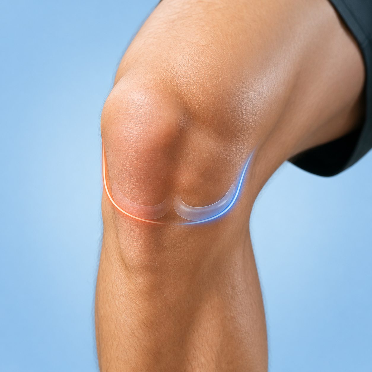
| Meniscus | Twist on a planted foot or lower-force twist in older tissue | Medial or lateral joint line | Deep bend pain, catching, reduced motion, locking | Clicking alone does not confirm a tear |
| Patellar or quadriceps tendon | Forceful jump, fall, or sudden loaded contraction | Below or above the kneecap | Difficulty actively straightening the knee | Ordinary tendinopathy usually develops differently |
| Bone or kneecap | Direct impact, fall, collision, or repetitive overload | Focal bony tenderness | Bearing weight or moving may be very difficult | Early stress injuries can have a normal X-ray |
Treat each row as a set of questions for an assessment, not a verdict. Multiple structures can be injured in one event. An ACL injury may occur with a meniscus tear or collateral-ligament injury, and a patellar dislocation may create early swelling that resembles another internal injury.
Start With the Event, Not the Loudest Symptom
The mechanism describes how force reached the knee. A planted foot followed by rotation differs from a direct blow to the front of a bent knee. A sideways collision differs from a repetitive increase in running. Reconstruct the two or three seconds before symptoms began: direction, speed, foot position, contact, landing, and whether the knee bent, twisted, or moved sideways.
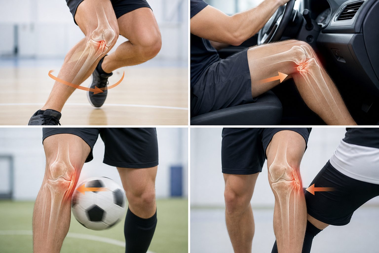
Memory after a sudden event is imperfect. Do not force a story to fit a diagram. If you cannot remember whether the knee moved inward or outward, say that. The later pattern may still be informative, including swelling timing, the most tender location, whether motion becomes blocked, and whether the knee feels stable during ordinary steps.
Swelling Timing: Minutes, Hours, or Days
Swelling that builds quickly after significant trauma can signal bleeding or a large inflammatory response inside the joint. ACL injury, patellar dislocation, fracture, and other internal injuries belong in that differential. Rapid swelling is a reason for prompt assessment, particularly when weight bearing or movement is limited.
Swelling that develops over several hours or during the next two to three days can occur with a meniscus injury or other joint irritation. The slower timeline does not make the problem trivial. Record whether the knee looks larger, feels full, loses its normal bend, or becomes harder to straighten. Compare both knees rather than relying on a vague sense of puffiness.
No visible swelling does not rule out a meaningful injury. Some collateral-ligament, tendon, cartilage, or bone-stress injuries produce limited swelling. A chart works best when swelling is combined with mechanism and function rather than treated as a required box.
ACL Injury Symptom Pattern
An ACL injury often follows a sudden stop, pivot, change of direction, awkward landing, or collision. People may describe a pop, rapid swelling, reduced movement, and a sense that the knee cannot be trusted. Pain may feel deep or spread across the knee rather than staying at one small surface point.
None of those symptoms is exclusive to the ACL. A pop can occur with meniscus injury, patellar dislocation, or a harmless joint sound. Giving way can reflect pain and quadriceps inhibition rather than a torn stabilizing ligament. Clinical tests, performed when pain and swelling allow, are what help evaluate ligament stability.
PCL Injury Symptom Pattern
The PCL limits backward movement of the shin relative to the thigh. A classic mechanism is a strong force against the front of a bent knee, such as a dashboard impact or a hard fall directly onto the bent knee. Symptoms may be felt deep in the knee or toward the back, with swelling and difficulty during deceleration, slopes, stairs, or kneeling.
Higher-energy trauma may injure more than the PCL. If the event involved a vehicle collision, major fall, visible deformity, numbness, or circulation change, do not use a symptom chart to decide that only one ligament is involved.
MCL and LCL Injury Symptoms
Collateral ligaments run along the sides of the knee. An inward collapse caused by a force from the outer side may stress the MCL on the inner knee. A force from the inner side may stress the LCL on the outer knee. Tenderness may follow the ligament rather than sitting precisely in the joint-line gap.
Inner and outer pain still overlap with meniscus, bone, tendon, and IT-band patterns. LCL-region trauma also deserves care because structures at the outer back corner of the knee can be involved. A clinician may compare side-to-side stability at different knee angles and assess the rest of the limb rather than pressing only where it hurts.
Meniscus Injury Symptoms
Meniscus injuries can follow a twist on a planted foot, especially with the knee bent. Degenerative tissue can become symptomatic after a much smaller twist, including rising from a chair. Common clues include medial or lateral joint-line tenderness, stiffness, swelling that may build over two or three days, pain with rotation or deep bending, catching, and reduced range.
True locking means the knee is physically blocked and cannot complete its normal movement, not simply that movement hurts. A displaced tear is one possible cause, but the symptom needs assessment. The meniscus pain location chart maps medial, lateral, and back joint-line patterns in detail.
Tendon and Bone Injury Patterns
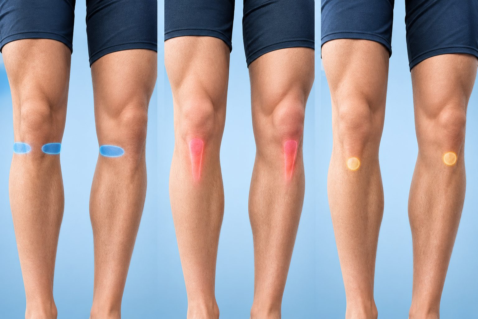
The quadriceps tendon sits above the kneecap and the patellar tendon runs from the kneecap to the upper shin. A sudden forceful contraction, jump, or fall may injure this extensor mechanism. A complete rupture can make an active straight-leg raise or knee extension impossible. That functional loss matters more than whether the person heard a pop.
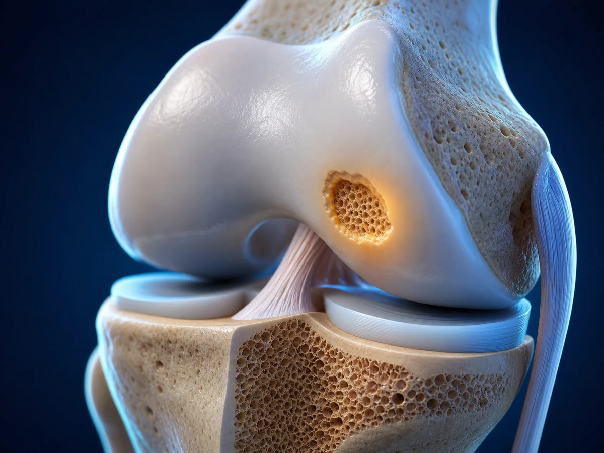
A fracture becomes more concerning after a direct impact or fall when there is severe focal tenderness, bruising, deformity, inability to bear weight, or inability to move the knee. Repetitive bone-stress injuries are different. They may begin with localized pain during activity, progress to walking or night pain, and can be missed on an early X-ray.
Pain Location Is One Layer of the Chart
- Deep or diffuse pain: can occur with major internal injury but is not specific to one ligament.
- Medial or lateral joint line: may fit a meniscus pattern, especially with twisting, delayed stiffness, catching, or locking.
- Inner or outer side band: may fit a collateral-ligament pattern after a sideways force.
- Directly below or above the kneecap: raises tendon questions, especially when active extension is weak or absent.
- One sharply tender bone point: increases concern for a bone injury after impact or repetitive overload.
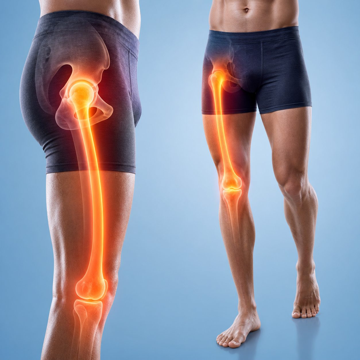
Surface pain can also be misleading. A swollen joint may hurt everywhere. Guarding changes where pressure is felt. Referred pain from the hip or spine can reach the knee without a new knee injury. If the event was unclear and hip movement also hurts, use the hip-to-knee referred pain map as a separate comparison.
What Function Can Tell You
Function does not grade the injury, but it can identify urgency. Can you take four ordinary steps without the knee collapsing? Can you fully straighten and comfortably bend it? Is motion limited by pain, swelling, or a hard mechanical block? Can you lift the straight leg from a bed? Does the foot feel warm and normal, or cold, numb, weak, or discolored?
Do not repeatedly test a painful knee with hopping, twisting, deep squats, or forceful ligament maneuvers. A home test can aggravate symptoms and a negative result may provide false reassurance. Record the ordinary functions that changed and let a clinician choose stress tests.
When X-Ray, MRI, or Ultrasound May Enter the Picture
Imaging should answer a clinical question. X-rays show bone and alignment and are often the first study after trauma when fracture criteria or examination findings justify them. MRI provides detail about ligaments, menisci, cartilage, bone marrow, and tendons, but not every knee injury needs immediate MRI. Ultrasound can assess selected superficial tendons and fluid collections dynamically, yet it is not a substitute for MRI when deep internal structures are the concern.
The knee X-ray vs MRI vs ultrasound guide explains what each test can show. The right sequence depends on trauma severity, examination, age, ability to bear weight, locking, extensor function, and whether the result would change treatment.
What to Record Before an Appointment
- The event: pivot, landing, side impact, fall, direct blow, or repetitive load increase.
- The first minute: pop, immediate pain, ability to continue, visible change, or loss of control.
- Swelling timeline: minutes, several hours, next morning, or none.
- Location: joint line, side band, kneecap, tendon, back of knee, or focal bone point.
- Function: weight bearing, bending, straightening, locking, buckling, stairs, and sleep.
- Changes since then: improving, stable, worsening, or repeatedly swelling after use.
A short timeline is more useful than a confident self-diagnosis. Bring information about previous knee injuries, surgery, medication, osteoporosis risk, and the activity you need to return to. Those details change the examination and recovery plan.
What a Knee Injury Chart Cannot Tell You
Symptom charts are best at organizing a history, not naming a torn structure. Pain location can spread after swelling develops. Guarding can make a stable knee feel weak. A person with a meaningful ligament or meniscus injury may still walk, while a painful bone bruise or minor sprain can make early walking very difficult. The apparent severity of one symptom therefore does not rank the seriousness of every possible injury.
Home maneuvers are limited for the same reason. Pulling on the shin, twisting the knee, or repeatedly forcing a deep bend can provoke several tissues and may worsen symptoms. Clinical tests are interpreted as a group and compared with the uninjured side. The examiner also checks hip and ankle movement, circulation, sensation, muscle function, and focal bone tenderness before deciding whether imaging is useful.
Age and context change the meaning of the chart. A non-contact pivot during sport, a dashboard impact, a fall onto the kneecap, and a low-energy twist in an older adult create different starting probabilities. Previous surgery, osteoporosis, inflammatory disease, anticoagulant medicine, and an artificial joint can also change urgency. Include those details when seeking advice rather than relying on the closest-looking row.
How Symptoms Can Change During the First 48 Hours
The first minutes do not always show the whole pattern. Adrenaline may make walking seem possible, then swelling and muscle inhibition can reduce motion over the next several hours. Meniscus-related stiffness may become clearer the next day. Bruising can take time to appear and may spread away from the injured structure. Record changes instead of repeatedly testing the knee at full effort.
Until the situation is clear, protect the knee from the movement that caused the event, use support if walking is unsafe, and follow appropriate medical guidance. Do not assume that a quieter second day proves the tissue is intact, and do not assume that bruising alone confirms a ligament tear. Function, examination, and the course over time need to agree.
When to Get Medical Help
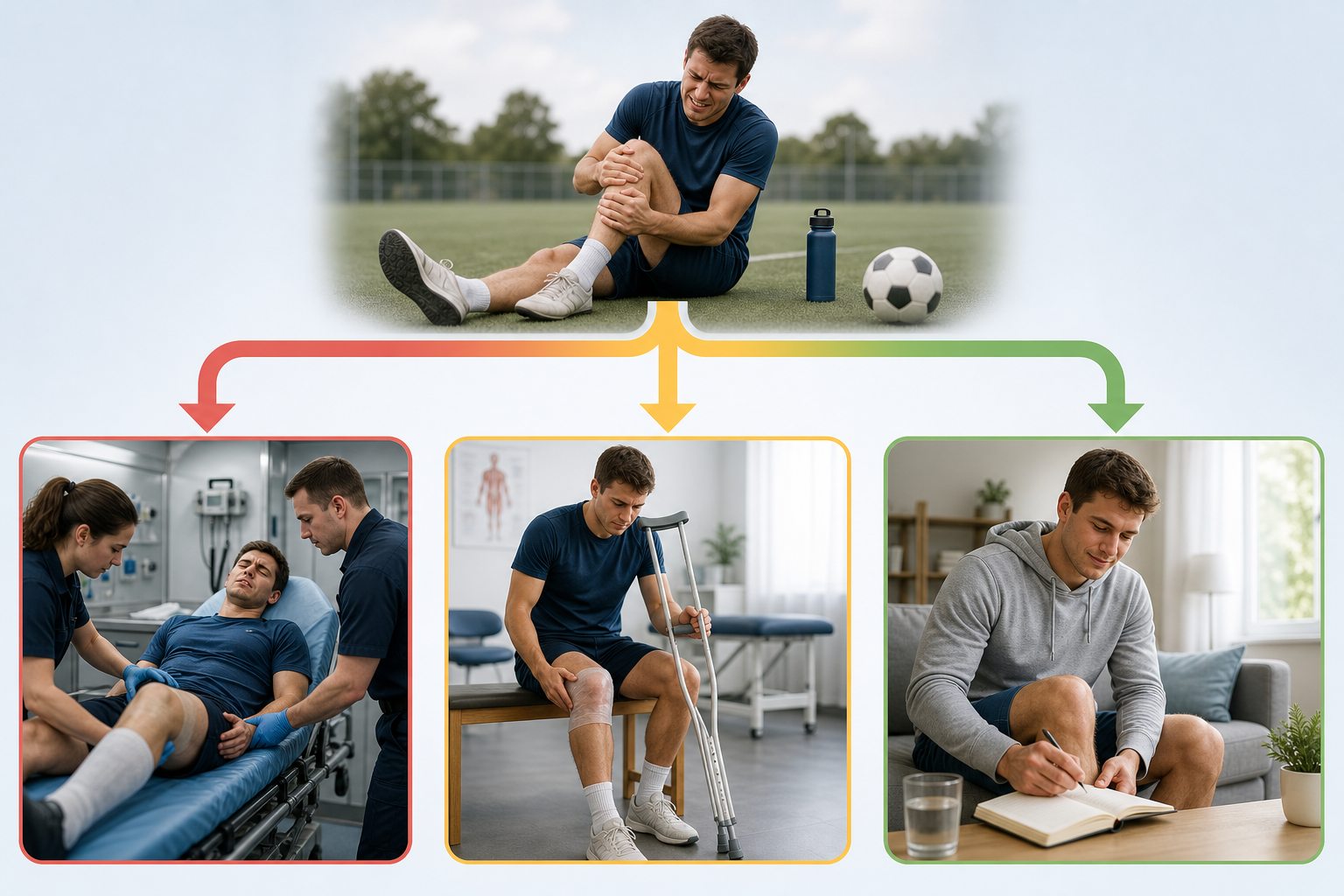
Seek urgent care for an obvious deformity, an open wound over a suspected fracture, a cold or discolored foot, new marked numbness or weakness, inability to bear weight after significant trauma, a physically locked knee, or severe rapidly increasing swelling. Arrange prompt assessment when swelling, instability, reduced motion, focal bone tenderness, or inability to actively straighten persists after an injury.
A backward-bending event has its own assessment questions in the hyperextended knee injury guide. When the event happened on a ski slope, the post-skiing knee pain guide adds equipment, terrain, and return-to-slope considerations.
The Bottom Line
A knee injury symptom chart should improve the description of an injury, not name it. Mechanism, swelling timing, pain location, stability, range, extensor function, and weight bearing are the useful layers. ACL, PCL, collateral-ligament, meniscus, tendon, and bone injuries share many symptoms and can occur together. Use the chart to prepare for the right level of assessment, then let examination and selective imaging establish what is actually injured.
Frequently Asked Questions
How can I tell what type of knee injury I have?
Start with the mechanism, swelling timing, exact pain location, stability, range of motion, and ability to bear weight. Those clues can prioritize possibilities, but they cannot confirm which tissue is injured. A clinician combines the history with examination and selects imaging only when it will change management.
Does rapid swelling mean an ACL tear?
Rapid swelling after a pivot, landing, or collision can occur with an ACL injury, but it is not specific. Patellar dislocation, fracture, and other internal injuries can also cause early swelling. A rapidly enlarging knee after trauma deserves prompt assessment rather than a home diagnosis.
What are the symptoms of a meniscus knee injury?
A meniscus pattern may include medial or lateral joint-line pain, stiffness or swelling that develops over hours to a few days, pain with twisting or deep bending, catching, reduced motion, or true locking. Many other knee problems can produce some of the same symptoms.
Can I still walk with a torn ligament or meniscus?
Yes. Some people can walk after a ligament or meniscus injury, especially before swelling and stiffness increase. Being able to walk does not rule out a meaningful injury. The mechanism, swelling, instability, locking, and later function still matter.
What knee injury causes the knee to give way?
Giving way may reflect ligament instability, pain-related muscle inhibition, patellar instability, or another problem. Repeated buckling after an injury deserves assessment because the sensation alone cannot identify the structure responsible.
When is a knee injury an emergency?
Seek urgent medical care for an obvious deformity, bone through the skin or a deep open wound, a cold or discolored foot, new marked numbness or weakness, inability to bear weight after significant trauma, a physically locked knee, or severe rapidly increasing swelling.