To understand meniscus tear symptoms, consider both where medial and lateral joint-line pain is felt and which other symptoms accompany it. Location alone is less informative than the full pattern of pain, swelling, movement, and injury history.
This guide stays deliberately narrow. The main knee pain location chart compares every knee region. The knee injury symptom chart compares ligament, meniscus, tendon, and bone patterns after trauma. Here, the focus is only the meniscus and the structures most commonly confused with it.
Meniscus Pain Location Chart
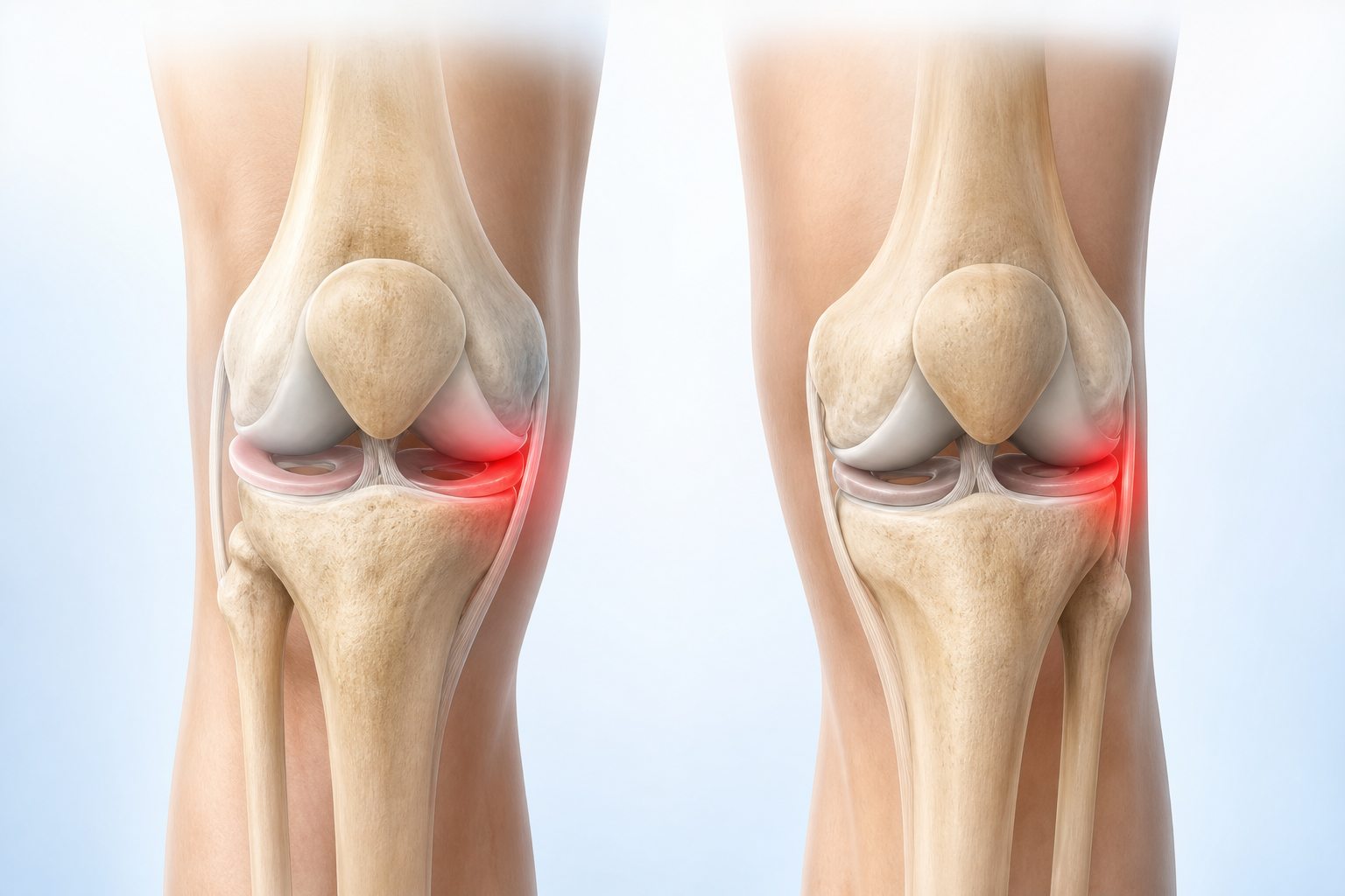
| Region | What people often feel | Clues that add context | Common alternatives |
|---|---|---|---|
| Medial joint line | Deep inner-knee pain or a narrow tender line | Twist, deep bend, delayed swelling, catching | MCL, pes anserine region, arthritis, bone |
| Lateral joint line | Deep outer-knee pain, sometimes toward the back | Pivot, rotation, loaded flexion, reduced motion | LCL, IT band, popliteus, arthritis, bone |
| Posteromedial or posterolateral line | Deep pain at the back corner of the joint | Full bend, rotation, swelling, blocked extension | Hamstring, calf, cyst, PCL, popliteus |
| Diffuse whole-knee ache | General stiffness or fullness rather than one line | Large effusion or several irritated tissues | Many joint, inflammatory, or traumatic causes |
What Is the Knee Joint Line?
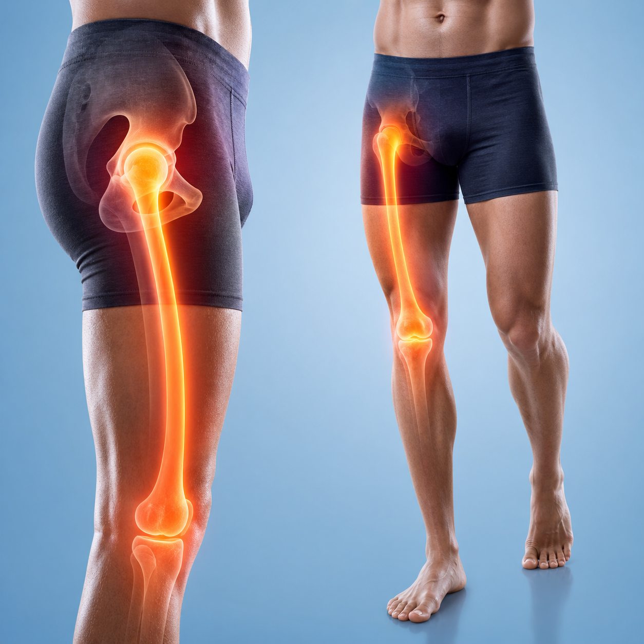
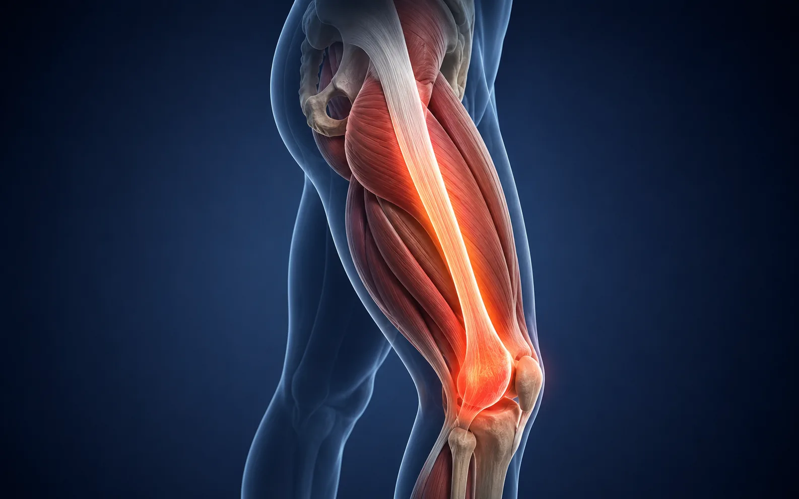
The joint line is the narrow gap where the femur meets the tibia. On a relaxed knee, a clinician can feel along the medial and lateral edges of that gap. The medial and lateral menisci are wedge-shaped fibrocartilage structures inside it. They help distribute load, contribute to joint stability, and move slightly as the knee bends and straightens.
Because the meniscus is inside the joint, people often describe the pain as deep rather than on the skin. The sore area may be easiest to find with the knee slightly bent. Tenderness exactly along the line is one examination clue, but it is not specific enough to diagnose a tear. Osteoarthritis and nearby ligament injuries can create the same finding.
Medial Meniscus Pain Location
The medial meniscus sits on the inner half of the knee. A symptomatic pattern may feel like a narrow deep ache along the inner joint line, sometimes extending toward the back-inner corner. A planted-foot twist, deep squat, kneeling turn, or getting up from a low chair can provoke it. The knee may become stiffer over several hours and lose some bend or extension.
Inner pain alone is not enough. The MCL lies on the inner side and can be injured by a sideways force. The pes anserine tendons and bursa sit lower on the inner shin, often below the joint line. Medial-compartment osteoarthritis can create joint-line aching without a new tear. A precise history keeps those patterns from being collapsed into one label.
A clinician may ask whether rotation or a deep loaded bend reproduces the familiar pain, then compare ligament stability, range, swelling, and tenderness above and below the line. The goal is not to make one test positive. It is to see whether several findings tell a consistent story.
Lateral Meniscus Pain Location
The lateral meniscus sits on the outer half of the knee. Symptoms may be felt at the lateral joint line or deeper toward the back-outer corner. Pivoting, cutting, deep bending, or rotating on the foot may provoke the area. Catching and a blocked feeling can occur, but a painless click is not proof of a tear.
Outer knee pain has important alternatives. The IT band is more superficial and often becomes sore with repeated running or cycling near the outer femoral epicondyle. The LCL follows the outer side and is more relevant after a sideways force. The popliteus region can hurt toward the posterolateral knee. The IT band pain location diagram helps separate a distance-related outer-thigh path from a joint-line pattern.
Can Meniscus Pain Be Felt Behind the Knee?
The back portions of both menisci sit toward the posterior joint line, so a meniscus problem can be experienced near a back corner, particularly with deep flexion. Yet pain directly behind the knee also has many other sources, including hamstring or calf tissue, a Baker's cyst, the popliteus region, ligament injury, and less commonly vascular or nerve problems.
A feeling of fullness with visible swelling is different from a narrow joint-line tenderness. Calf swelling, redness, unusual warmth, or breathing difficulty needs urgent medical consideration rather than another meniscus self-test. The back-of-knee pain guide compares those posterior patterns.
Movements That Add Meaning to the Location
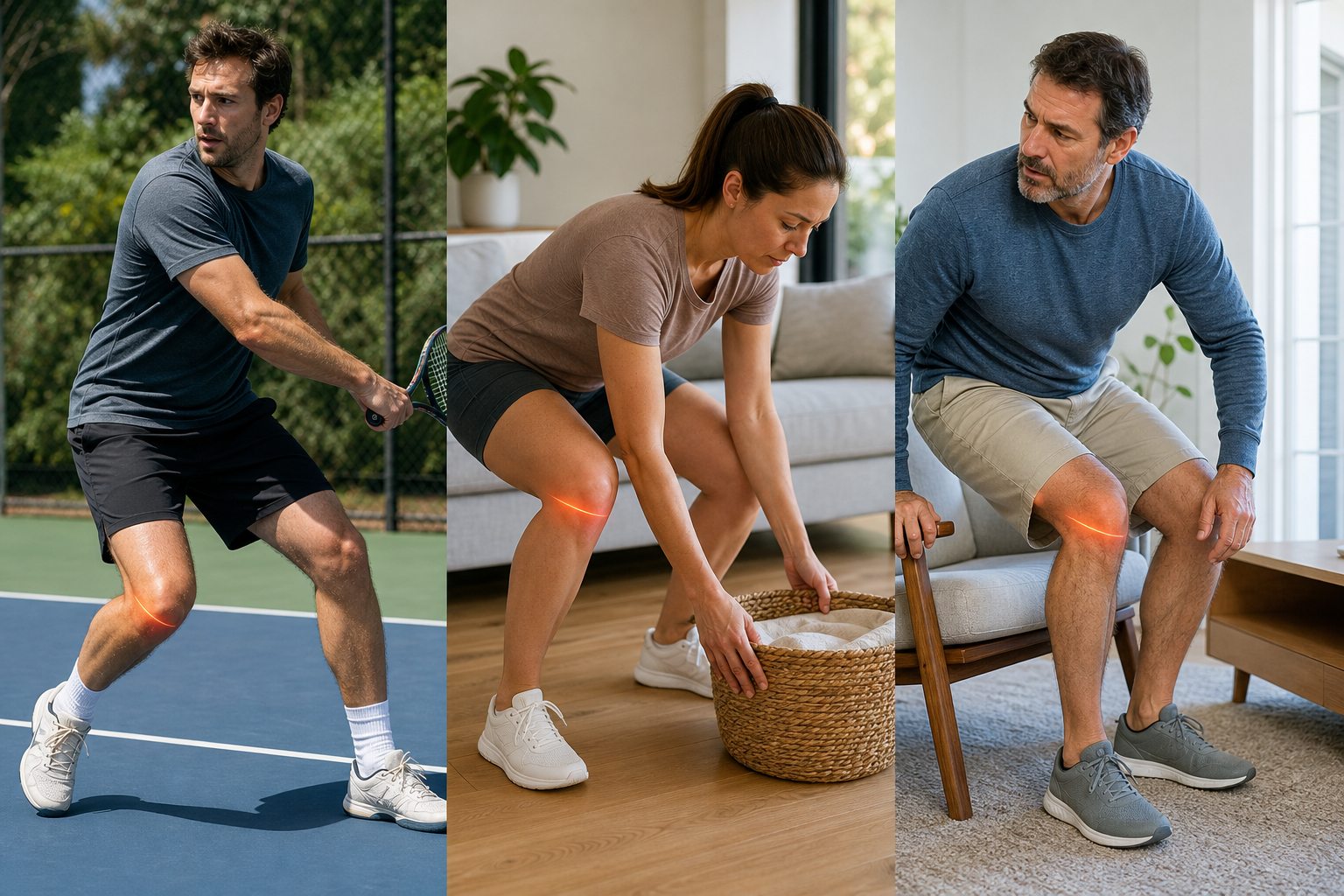
Twisting on a Planted Foot
Acute tears commonly occur when the foot remains planted while the body turns. Direction, knee bend, speed, and contact all affect what else may be injured. A meniscus tear can occur alongside an ACL injury, so early swelling or instability should not be attributed to cartilage alone.
Deep Bending and Squatting
Deep flexion moves and loads the menisci, but it also raises demand at the kneecap joint and tendons. Pain at the bottom of a squat therefore does not identify the meniscus. Joint-line location, a preceding twist, swelling, catching, and a blocked range make the pattern more specific.
Rising From a Chair
Age-related meniscal tissue can become symptomatic after a lower-force twist, including turning while rising. That does not mean every older adult with start-up knee pain has a tear requiring surgery. Osteoarthritis, stiffness after sitting, muscle capacity, and seat height can produce the same functional complaint.
Clicking, Catching, and True Locking Are Not the Same
Clicking is a sound or sensation and is common in knees with and without pain. Catching is a brief interruption or snag during motion. True locking means the knee is physically blocked and cannot complete its normal bend or straightening. Painful guarding may feel like locking, but the distinction matters because a displaced meniscal fragment is one reason a knee can become mechanically blocked.
Do not repeatedly force a locked knee or have another person twist it. Note the angle where it stops and arrange prompt assessment. If the main symptom is a painless sound during walking, bending, or squatting, our knee clicking guide is the more appropriate page.
Meniscus Pain vs Common Look-Alikes
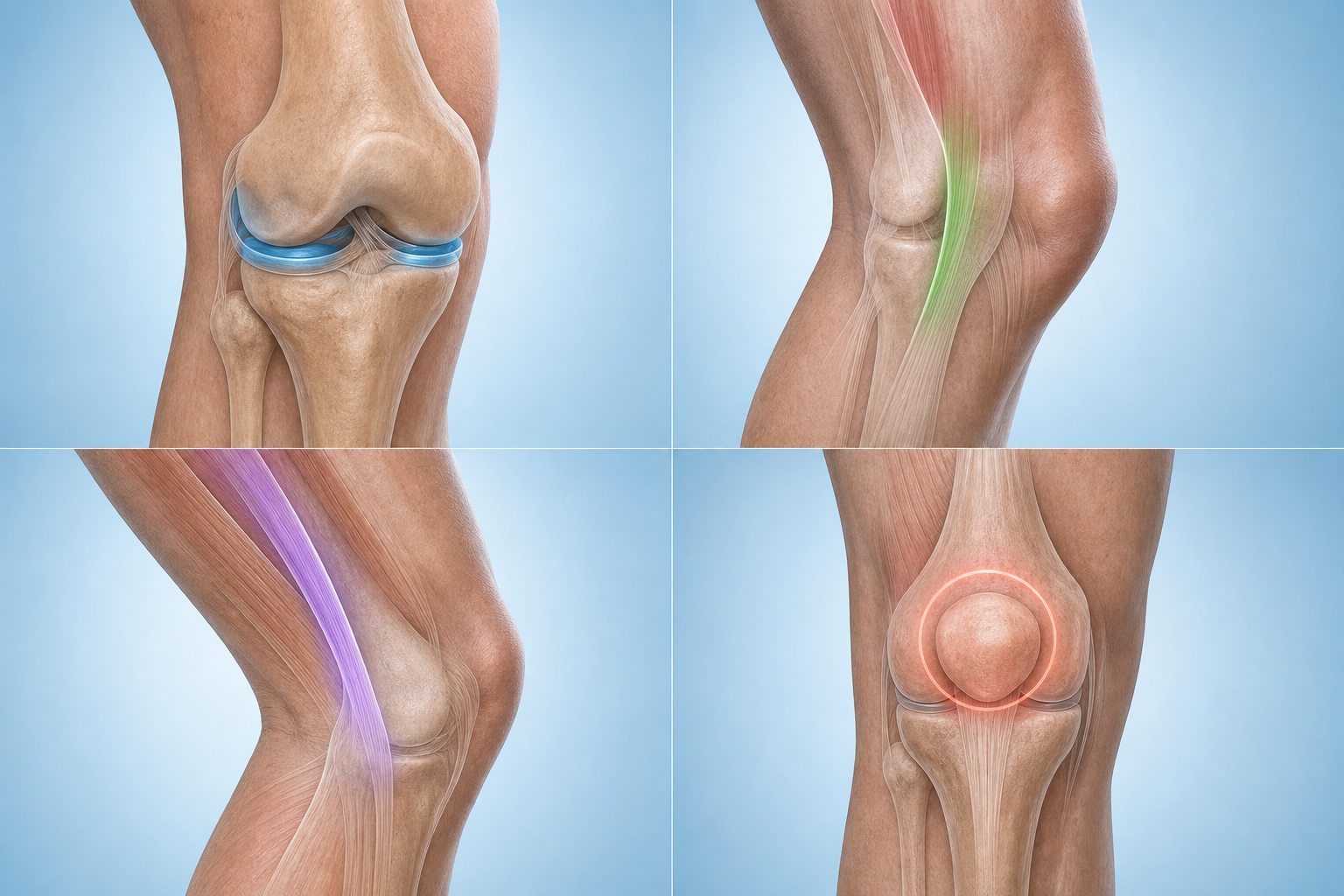
| Pattern | Location | Typical trigger | Clue against a simple meniscus label |
|---|---|---|---|
| MCL | Inner side, often a line broader than the joint gap | Force that pushes the knee inward | Pain follows ligament stress rather than rotation alone |
| Pes anserine region | Inner upper shin, below the joint line | Stairs, hills, repeated flexion | Tenderness is lower and more superficial |
| IT band | Outer knee, often with lateral thigh awareness | Running or cycling volume | Distance and repetition dominate the story |
| Patellofemoral | Around or behind the kneecap | Stairs, squats, running, prolonged sitting | Pain is anterior rather than at a joint line |
| Osteoarthritis | Can include the medial or lateral joint line | Load, stiffness after rest, fluctuating activity | No single twist may explain the longer pattern |
Acute vs Degenerative Meniscus Tears
An acute traumatic tear often follows a memorable pivot or loaded twist. Degenerative tears develop in tissue changed by age and cumulative load, and the triggering movement may be minor or absent. MRI can reveal a degenerative tear that was not responsible for every symptom, especially when osteoarthritis is also present.
This is why treatment is not chosen from the scan phrase alone. In trials involving middle-aged adults with degenerative meniscal tears, structured exercise therapy has often produced outcomes comparable with arthroscopic partial meniscectomy for many participants. Those findings do not apply automatically to a newly locked knee, acute repairable tear, major associated injury, or every individual.
The practical question is whether symptoms and function are improving with an appropriate non-surgical plan, or whether mechanical blockage, instability, persistent swelling, or activity needs justify a surgical opinion. Age by itself does not answer that question.
What a Clinician Checks
Assessment begins with mechanism and timeline. The clinician may inspect swelling, measure range, palpate both joint lines, assess ligaments, observe walking, and use carefully selected rotation or flexion tests. No single maneuver is perfect. A combination of findings is interpreted with the whole story.
X-rays do not show a meniscus tear, but they can identify fracture, alignment, and osteoarthritis. MRI shows the menisci and associated soft tissues in detail. It is most useful when the result will change management, the diagnosis remains uncertain, or surgery is being considered. The knee imaging comparison explains why an immediate MRI is not always the first step.
What to Do While Waiting for Assessment
Protect the knee from the movement that caused sharp pain, particularly pivoting and forced deep flexion. That does not always require complete rest. Comfortable range-of-motion work and ordinary walking may be reasonable when the knee is stable, not locked, and symptoms are mild. A clinician can adapt loading after a major injury or when diagnosis is unclear.
Track swelling, range, weight-bearing tolerance, sleep, and whether the knee catches or gives way. Avoid repeating internet tests until one hurts. Do not use a sleeve, strap, or pain relief as permission to return immediately to cutting sport when the knee remains swollen or unstable.
Why a Meniscus Pain Diagram Is Easy to Misread
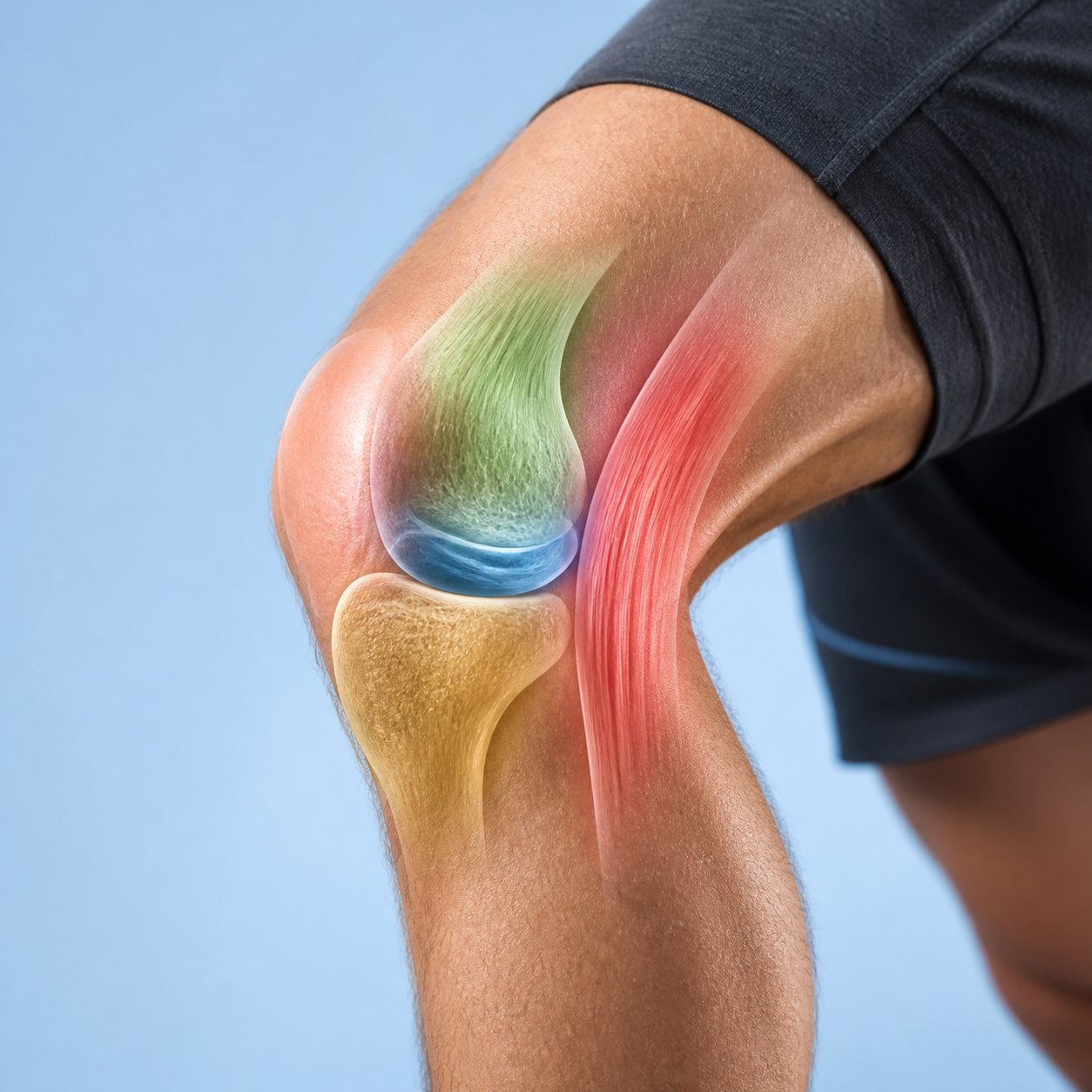
A diagram draws tidy colored zones, but real symptoms do not respect a border. The medial meniscus sits close to the MCL, pes anserine tendons, and the inner edge of the kneecap. The lateral joint line sits near the LCL, popliteus, proximal tibiofibular joint, and structures associated with the IT band. Swelling can make the whole knee feel full, and a person may point to the front even when the most useful tenderness is at the joint line.
Location becomes more informative when paired with a repeatable movement. Inner joint-line pain after a twist, discomfort with deep flexion, and a new loss of extension form a different cluster from broad aching after a long walk. Likewise, a painless click during a squat is not the same as a sharp catch that physically stops the knee. Use the chart to describe the cluster, not to self-confirm a tear.
Search interest in meniscus tear symptoms and treatment is much higher than interest in the chart phrase itself. That can encourage an early jump from pain location to surgery. Many meniscus findings are managed without immediate surgery, especially when there is no true locking or major associated injury. Management depends on age, tear pattern, symptoms, activity goals, osteoarthritis, and response to a structured plan.
Questions to Ask About the Next Step
- Is the knee truly locked? A knee that cannot reach its usual extension is different from one that is painful but can still move.
- Was there a clear injury? A pivot with rapid swelling carries a different context from gradual joint-line aching.
- Would imaging change management? MRI can show meniscal tissue, but an image finding must still match the symptoms and examination.
- Is osteoarthritis part of the picture? Degenerative meniscus changes and joint arthritis commonly coexist.
- What function is the priority? Walking, kneeling, work, running, and field sport require different progressions.
- What should improve first? Swelling, extension, quadriceps control, confidence, and activity tolerance may recover at different rates.
If imaging is being considered, the knee X-ray, MRI, and ultrasound comparison explains what each test can and cannot show. An MRI report should answer a clinical question, not replace one.
When to Get Medical Help
Arrange prompt assessment for a physically locked knee, significant swelling after a twist, inability to regain normal motion, repeated giving way, or pain that prevents ordinary weight bearing. Seek urgent care after major trauma when the knee is deformed, you cannot bear weight, the foot becomes cold or discolored, or marked numbness or weakness appears.
The Bottom Line
Meniscus pain is commonly mapped to the medial or lateral joint line and may extend toward a back corner of the knee. The map becomes more useful when a planted-foot twist, delayed swelling, deep-bend pain, catching, locking, or reduced motion is present. It still cannot confirm a tear or decide treatment. Compare the location with nearby structures, record the timeline, and use a clinical examination to determine whether imaging or a specific rehabilitation pathway is needed.
Frequently Asked Questions
Where is meniscus pain usually located?
Meniscus-related pain is often felt along the medial or lateral joint line, the gap between the thighbone and shinbone. It may feel deep and can extend toward the back of the joint. Location is only one clue because ligaments, tendons, arthritis, and bone can hurt nearby.
Is inner knee pain always a medial meniscus tear?
No. Inner knee pain can also come from the MCL, pes anserine tendons or bursa, osteoarthritis, bone, or referred pain. A twist, delayed swelling, joint-line tenderness, catching, locking, and reduced motion may raise suspicion, but an examination is needed.
Can lateral meniscus pain be mistaken for IT band pain?
Yes. Lateral meniscus pain is usually at the outer joint line and may follow twisting or deep bending. IT band pain is often more superficial near the outer femoral epicondyle and is strongly linked to repeated running or cycling. The patterns overlap and should not be diagnosed from one sore point.
Can you walk with a torn meniscus?
Many people can still walk after a meniscus tear, especially before swelling and stiffness increase. Walking ability does not rule out a tear. A locked knee, worsening swelling, repeated giving way, or inability to regain motion needs assessment.
Does meniscus pain always cause swelling?
No. Some meniscus injuries cause little visible swelling. Others become stiff and swollen over hours or the next two to three days. Absence of swelling does not prove the meniscus is normal, and swelling itself is not specific to a tear.
Will a meniscus tear heal on its own?
Healing potential depends on tear location, blood supply, pattern, stability, age, associated injury, symptoms, and activity needs. Some tears improve with non-surgical care, while a displaced tear causing locking may need different management. Imaging findings alone do not decide treatment.